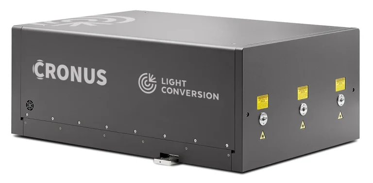
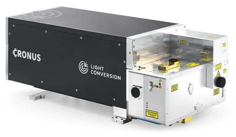
Light Conversion 的产品组合包括专为显微镜设计的飞秒激光器 CRONUS-2P 和 CRONUS-3P。
这些激光器适用于功能性神经成像、光遗传学和深层成像,采用中等重复率的三光子激发和快速高重复率的双光子成像,以及使用高功率激光器进行的宽视场和全息激发。请参阅下方 CRONUS 系列的对比表。有关非线性显微镜可用激光器的完整列表及其先进应用示例,请参阅最新宣传册。
| 产品 | 输出 | 输出功率 | 重复频率 | 脉宽 |
|---|---|---|---|---|
| 680 – 960 nm, 940 – 1300 nm, and 1025 nm 1) | >3 W @ 920 nm >2.5 W @ 1100 nm >2.5 W @ 1025 nm | 77 ± 1 MHz | < 160 fs | |
| 1250 – 1800 nm 2) 3) | > 1.1 W @ 1300 nm 4) > 0.8 W @ 1700 nm 4) | 高达 2 MHz | < 50 fs @ 1300 nm < 65 fs @ 1700 nm | |
| 1030 nm | 40 W (高达 120 W) | 高达 10 MHz | < 250 fs | |
| 1030 nm | 4 W (SP) (高达 20 W) | 10, 40, 或 80 MHz | < 50 fs (SP) |
- 最多可实现三个同时同步的输出,用于多光束激发。对于固定波长激光器,请参阅 FLINT。
- 对于双输出,请参考 ORPHEUS-F 配置中的 ORPHEUS-TWINS。
- 可提供具有额外 920 nm 输出的替代配置, 详情咨询 sales@lightcon.com。
- 在 1 MHz 的重复率下。可选择更低的重复率和更高的脉冲能量。
- 表示为 NRMSD (归一化均方根偏差)。
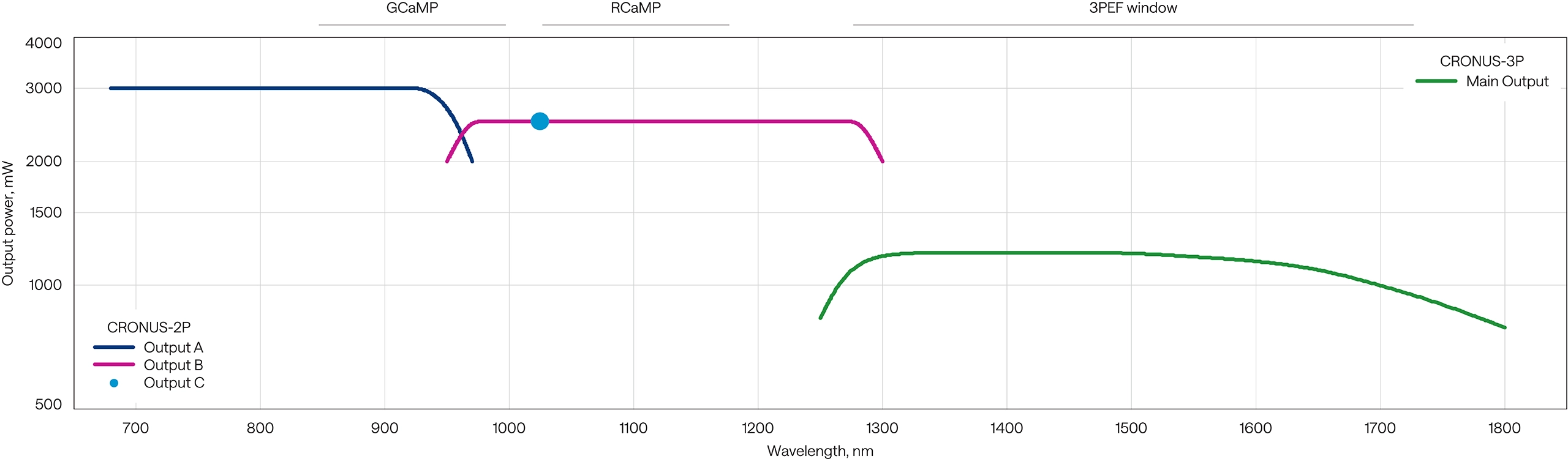
CRONUS-2P 和 CRONUS-3P 的调谐曲线
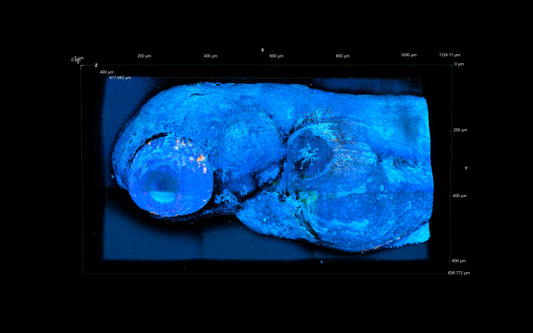
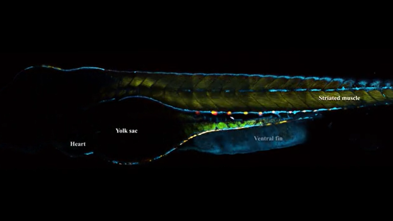
Multimodal 3D imaging of a live zebrafish at 1300 nm excitation using CRONUS-3P.
3PEF – green, trans SHG – yellow, epi THG – dark blue,
trans THG – cyan.
Courtesy of Luigi Bonacina group, University of Geneva.
Multimodal 3D imaging of a live zebrafish at 1300 nm excitation using CRONUS-3P.
3PEF – green, trans SHG – yellow, epi THG – dark blue,
trans THG – cyan.
Courtesy of Luigi Bonacina group, University of Geneva (2024).
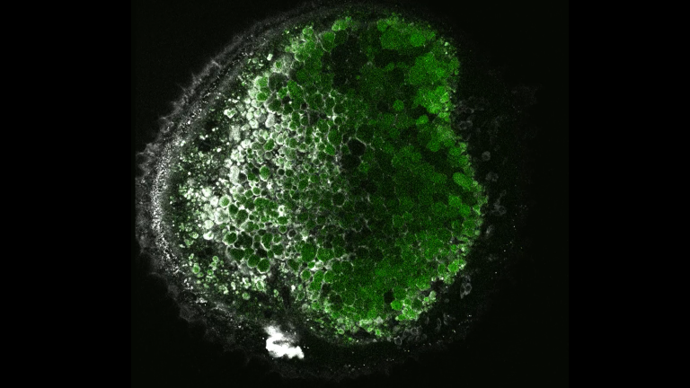
Multimodal 3D imaging of a live zebrafish yolk sac at 1300 nm 3P excitation using CRONUS-3P.
3PEF – green, transmitted THG – white.
Courtesy of Luigi Bonacina group, University of Geneva (2024).
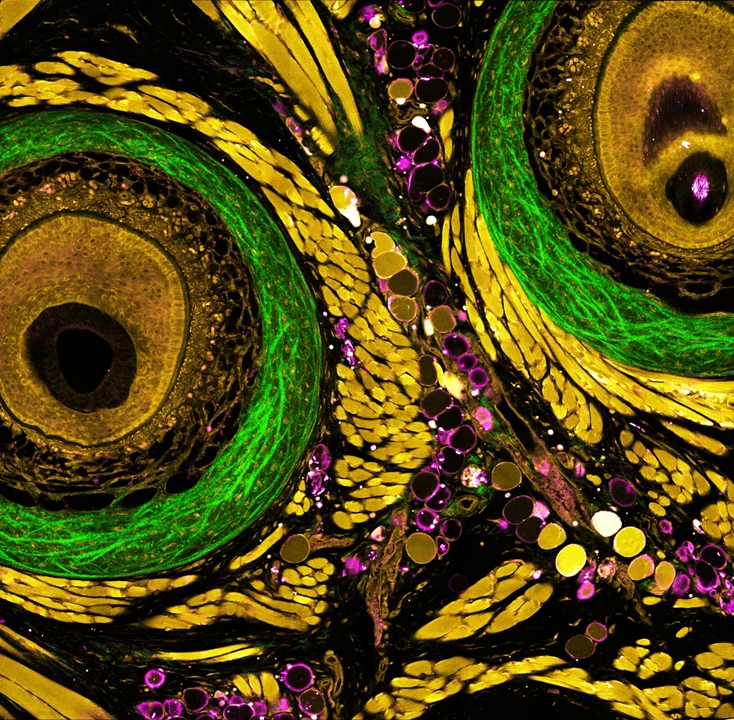
The microenvironment of an intact mouse whisker pad, revealed through multiplex label-free nonlinear microscopy using CRONUS-3P, illuminated at 1300 nm.
Courtesy of Kunzan Liu, Sixian You laboratory, Massachusetts Institute of Technology.
2P light-sheet imaging of the atrium in an adult mouse, acquired using the CARBIDE femtosecond laser with I-OPA.
Courtesy of Bliq Photonics.

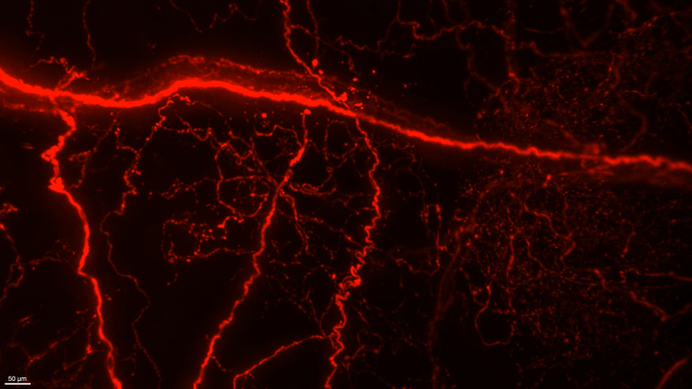
Mouse olfactory bulb with inhibitory cells labelled with GCaMP8s. Anatomical Z-stack imaged in 3P at 1300 nm down to 650 µm, 20 µm per step.
Source: Fred Marbach, Andreas Schaefer lab, The Francis Crick Institute, unpublished 2025 data.
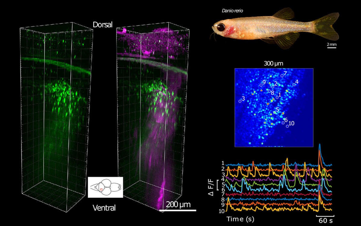
Functional three-photon neuroimaging of a zebrafish using an OEM OPA in the ORPHEUS-F configuration.
Courtesy of Chris Xu and Joseph R. Fetcho groups, Cornell University.
DOI: 10.1038/s41592-020-0819-7
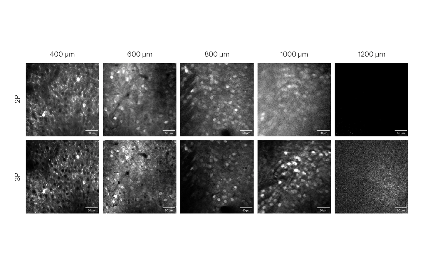
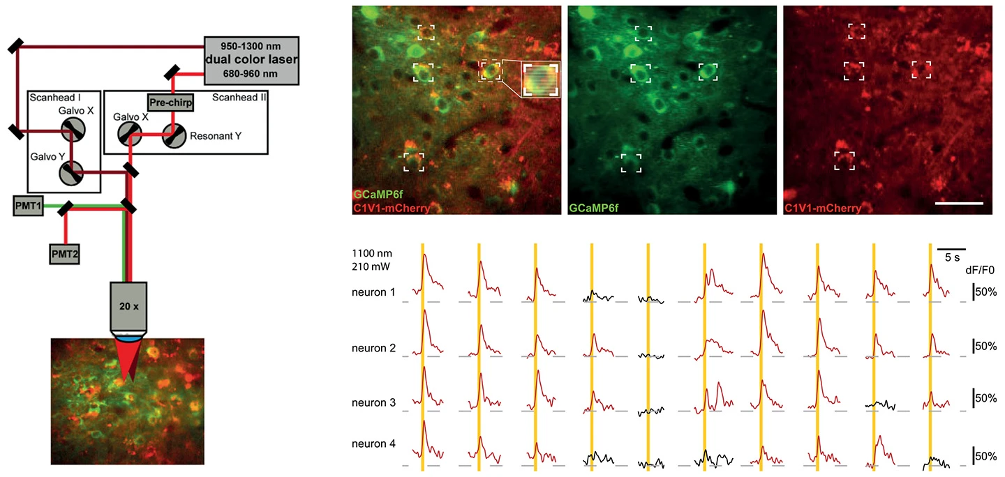
Comparison of in vivo 2P and 3P calcium imaging of mouse visual cortex GCaMP neurons on a Thorlabs Bergamo II microscope using typical 2P laser and Light Conversion CRONUS-3P (3P) laser at 920 nm and 1300 nm, respectively.
Courtesy of CSHL ISFNS 2024 school organizers and Willis Broden Jr. and Sergey Matveev (Thorlabs).
Adult zebrafish heart ventricle section, imaged using the FLINT femtosecond oscillator.
Samples courtesy of Justas Lazutka at the Vilnius University Life Sciences Center. Nonlinear imaging courtesy of the Barzda group at the Vilnius University Department of Physics.
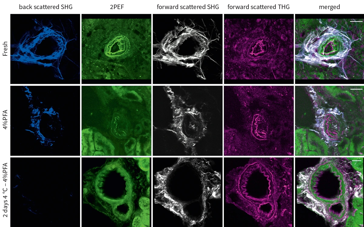
SHG signals from collagen, 2PEF, and THG signals from elastin in vibratome sections of mouse kidney after different treatments, acquired using CRONUS-2P.
Courtesy of Frauke Alves and Fernanda Ramos-Gomes, Max-Planck Institute for Multidisciplinary Sciences, Germany.
DOI: 10.1364/BOE.488453
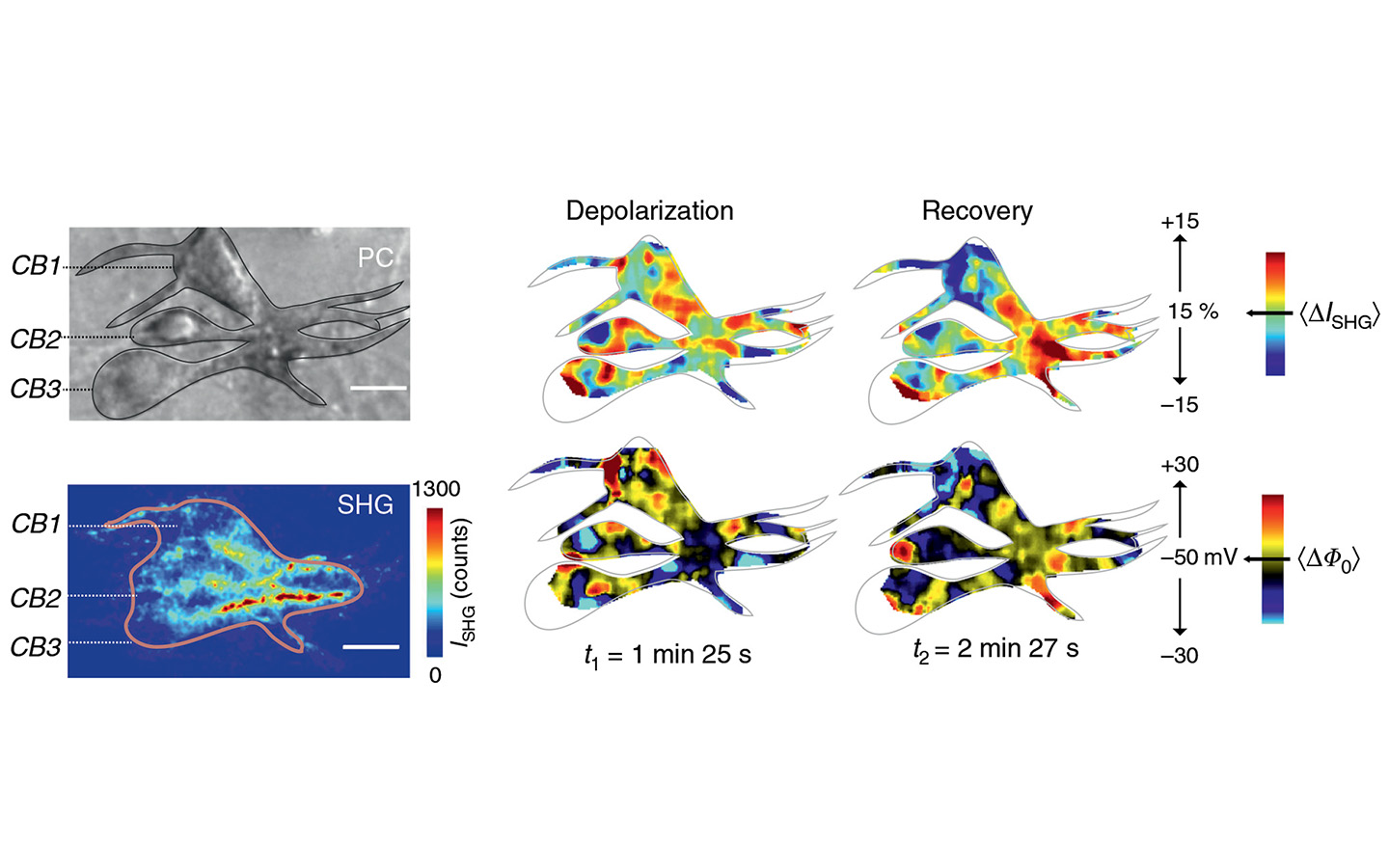
Widefield SHG neuroimaging of neuronal membrane potentials and ion efflux by means of water, acquired using the PHAROS femtosecond laser.
Courtesy of Sylvie Roke group, École Polytechnique Fédérale de Lausanne.
DOI: 10.1038/s41467-018-07713-w
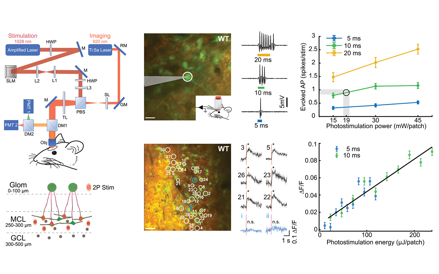
Holographic 2P optogenetic stimulation of mouse olfactory bulb neurons using the PHAROS femtosecond laser.
Courtesy of Shy Shoham and Dmitry Rinberg groups, New York University.
DOI: 10.1016/j.neuron.2020.07.034
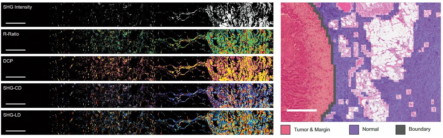
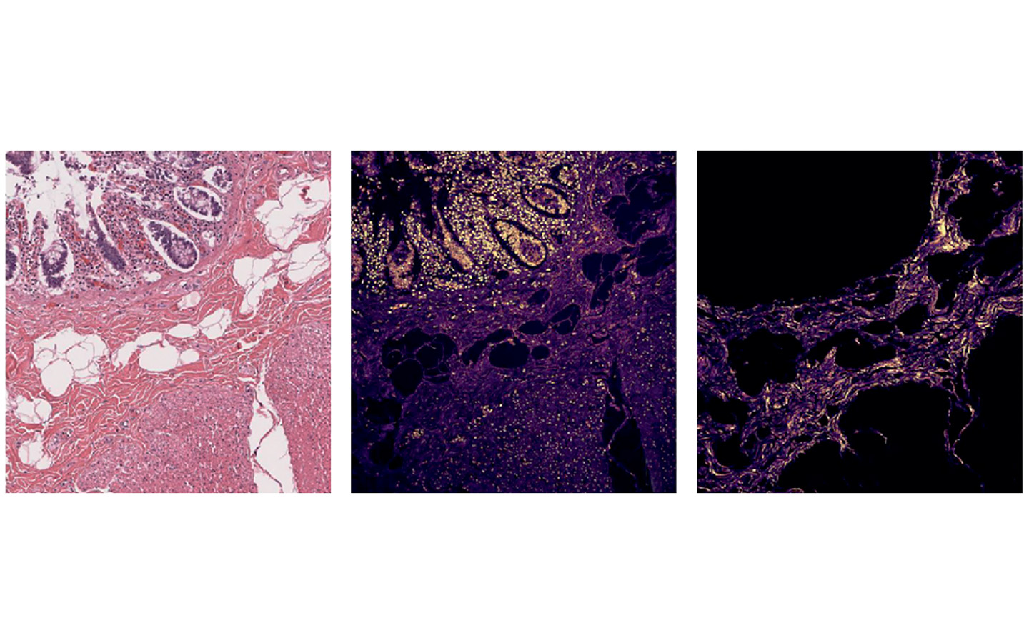
Large-area wide-field polarization-resolved SHG microscopy of a human lung tissue tumor margin using the PHAROS femtosecond laser.
Courtesy of Virginijus Barzda group, University of Toronto, and Brian C. Wilson group, Princess Margaret Cancer Centre.
DOI: 10.1038/s41598-022-24973-1

3P microscopy with adaptive optics for focus sensing and shaping to compensate for both aberrations and scattering. ORPHEUS-F excitation at 1300 nm enabled imaging up to 1.1 mm below the pia within the intact brain.
Courtesy of Jianan Y. Qu group, the Hong Kong University of Science and Technology.
DOI: 10.1038/s41587-022-01343-w
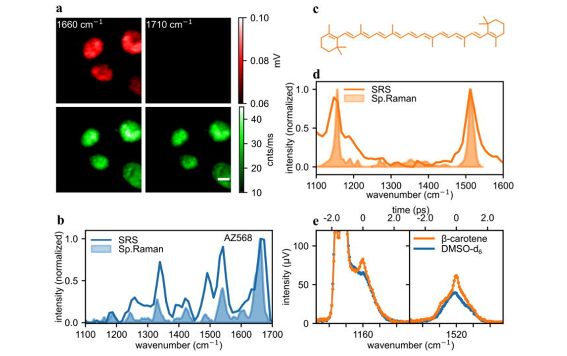
(a) Imaging of nuclei of AZ568 stained, fixed HeLa cells with SRS (red) and fluorescence (green). (b) SRS and spontaneous Raman spectrum (at 488 nm) of 1 mM solution of AZ568 in DMSO-d6. (c) Structure of β-carotene. (d) β-Carotene spectrum recorded with SRS and spontaneous Raman. (e) Spectral focusing spectrum of β-carotene solution in DMSO-d6. The images were recorded using CRONUS-2P.
DOI: 10.1063/5.0171725
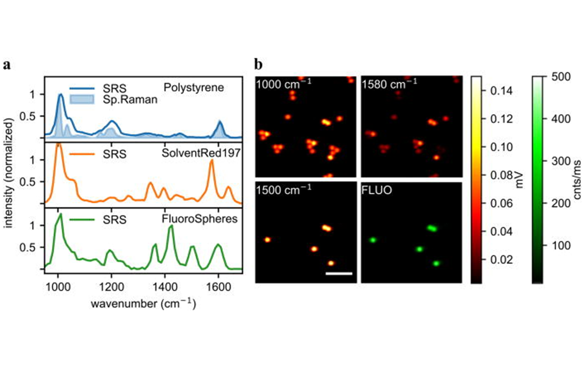
(a) Spontaneous Raman and SRS spectra of polymeric materials, normalized with the molecule of interest maximum peak. (b) SRS images of dyed and un-dyed PS beads. Images were recorded using CRONUS-2P.
DOI: 10.1063/5.0171725
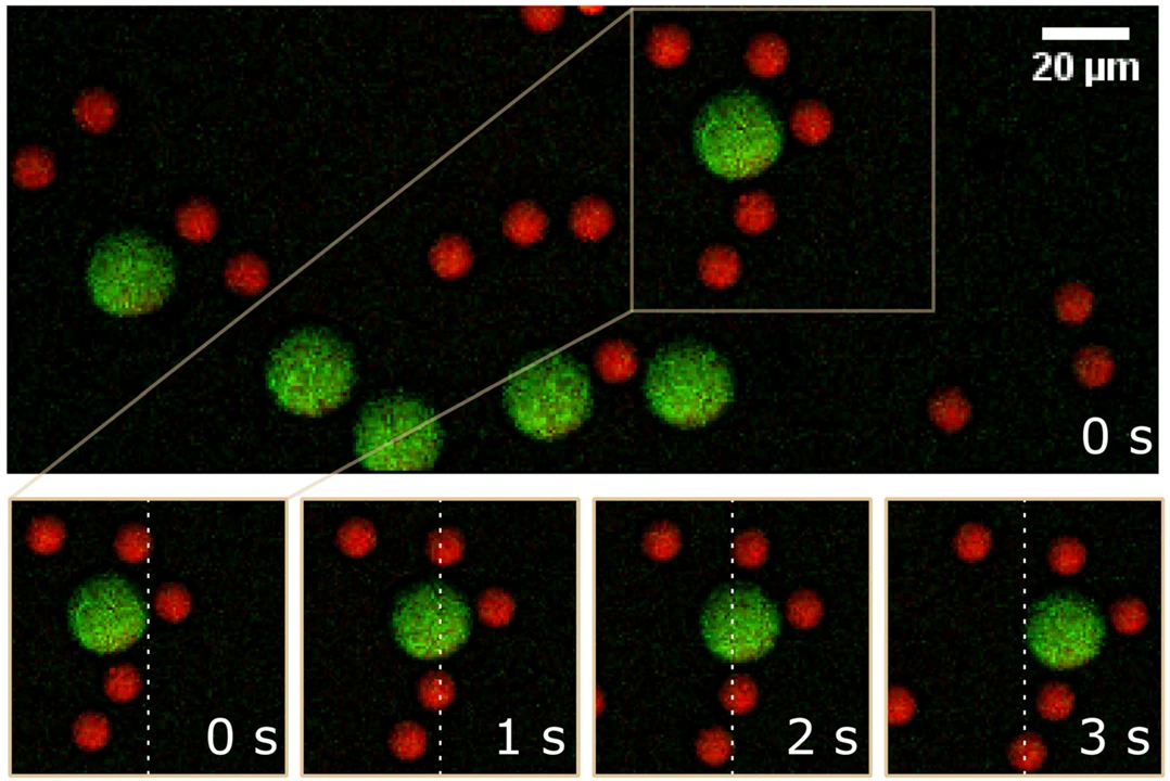
CARS images of flowing PS and PMMA beads, acquired using CRONUS-2P. The bottom images show a time course of a selected area as indicated with 1 s intervals to exemplify the flow. The white dashed line is for motion reference.
DOI: 10.1002/jrs.6671
2P optogenetic stimulation of individual neurons using CRONUS-2P.
Courtesy of Albrecht Stroh group, University Medical Center Mainz and Leibniz Institute for Resilience Research.
DOI: 10.1016/j.isci.2021.102184
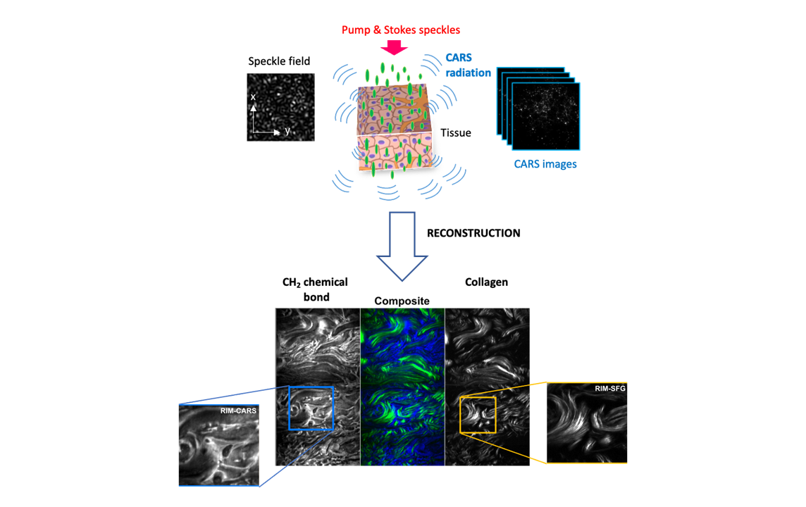
RIM-CARS and RIM-SFG imaging of molecular chemical bonds with a wide-field camera, using PHAROS and ORPHEUS-HP.
DOI: 10.1038/s41566-023-01294-x
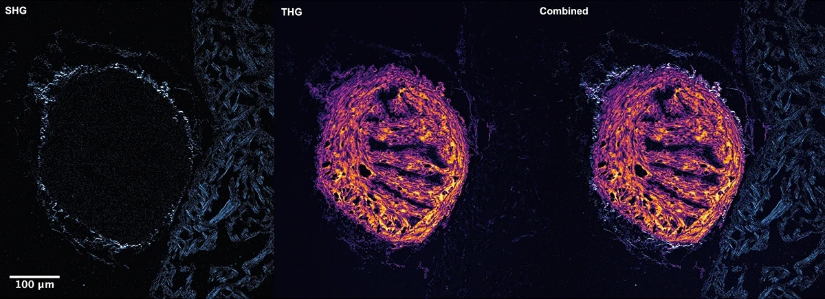
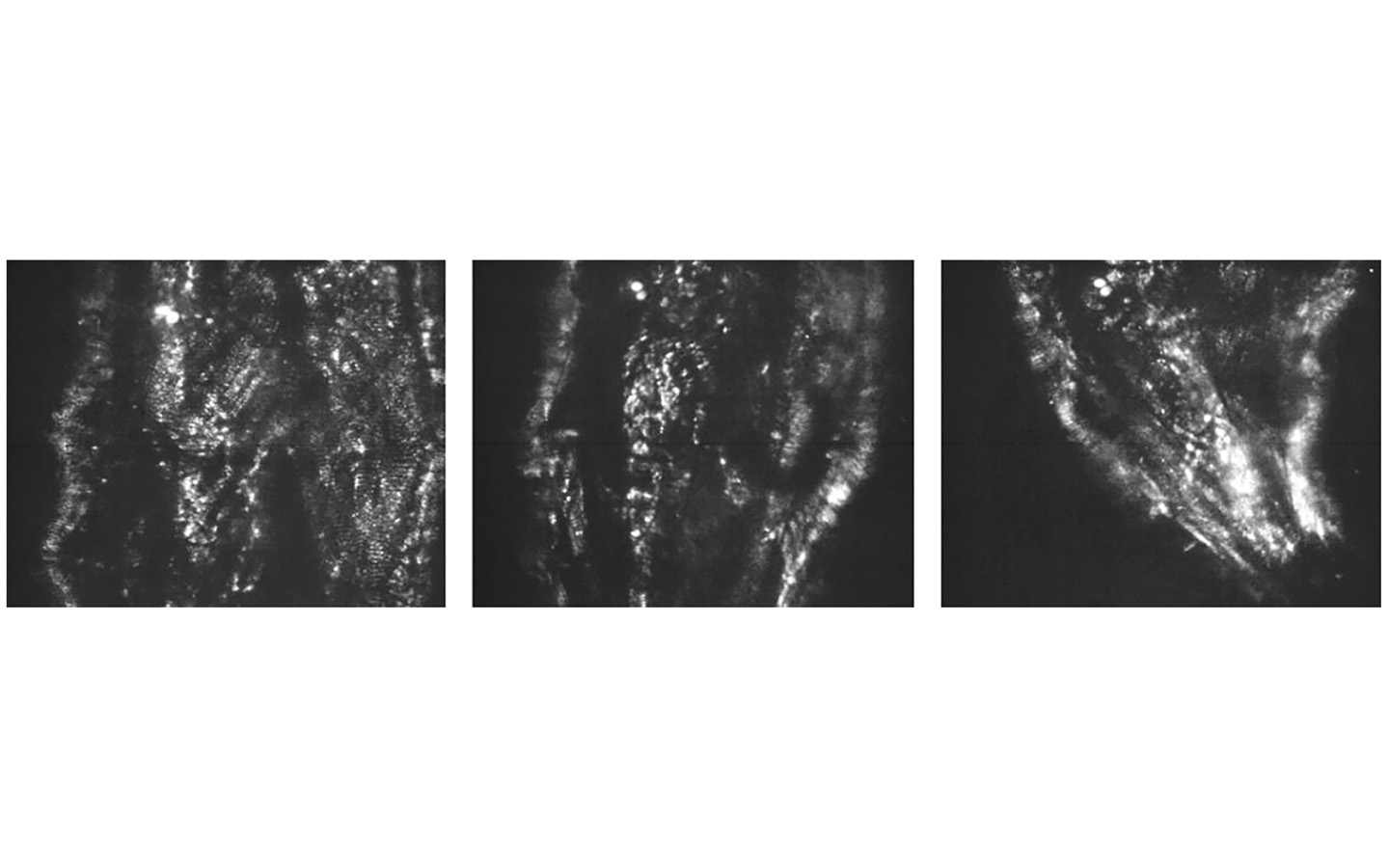
SHG and THG images of H&E-stained colon using the FLINT femtosecond oscillator.
Courtesy of Virgis Barzda group, Vilnius University.
Label-free in vivo widefield SHG imaging of a fruit fly larva using the PHAROS femtosecond laser.
Courtesy of Virgis Barzda group, University of Toronto.