
Nonlinear microscopy is a powerful technique for imaging inside living organisms with a submicrometer resolution at millimeter depths. In conjunction with genetically-encoded calcium indicators and opsins, multiphoton fluorescence (MPEF) microscopy has revolutionized neuroimaging and is becoming a standard tool in neuroscience, while label-free methods such as second- and third-harmonic generation (SHG and THG), and coherent anti-Stokes and stimulated Raman scattering (CARS and SRS) have been developed into ultrasensitive structural and chemical imaging techniques.
The nonlinear optical processes of nonlinear microscopy require high light intensities, which can be reached at low average power when ultrashort light pulses are tightly focused. This feature is exploited to provide optical sectioning and to improve imaging contrast deep inside scattering tissues. Multiphoton excitation occurs when two or more photons simultaneously pass in the vicinity of a molecule, and their combined energy is used for excitation leading to fluorescence. The simultaneous arrival of several photons can also result in a harmonic generation – radiation at double or triple of the excitation laser frequency. Harmonic generation is an intrinsic label-free contrast determined by and used to characterize the molecular order and homogeneity of the sample.
Higher-order, three- and four-photon-excited (3PEF and 4PEF), fluorescence microscopy deserves special attention as it enables imaging at depths that cannot be achieved with conventional microscopy techniques, especially in strongly scattering samples such as the brain. Most importantly, modern laser sources can operate at the required repetition rates and deliver the necessary pulse energy for real-time functional brain imaging at biologically-relevant depths.
CRONUS-3P is an OPA-based laser source that was developed specifically for nonlinear microscopy. It provides µJ-level sub-85 fs pulses at repetition rates of up to 2 MHz and tunable from 1.25 µm to 1.8 µm, thus covering the biological transparency windows at 1.3 µm and 1.7 µm for 3PEF microscopy. CRONUS-3P has integrated group delay dispersion (GDD) control, ensuring optimal pulse duration at the sample, and optional automated beam steering to guarantee laser pointing stability.
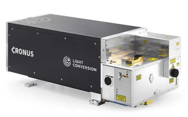
- 用于深度成像的高单脉冲能量
- 用于 3P 成像的 1250 – 1800 nm 调谐范围
- 低至 50 fs 的脉宽,高峰值功率
- 自动波长和 GDD 控制,以获得最佳信号
- 市场领先的脉冲间能量稳定性
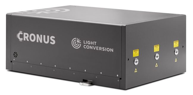
- 高重复频率下的瓦特级功率输出,用于快速成像
- 两个可调谐和一个固定的输出,用于同时进行多波束激发
- 自动化GDD控制,可在样品处获得最短的脉冲
- 工业级设计,具有高的输出功率和光束稳定性
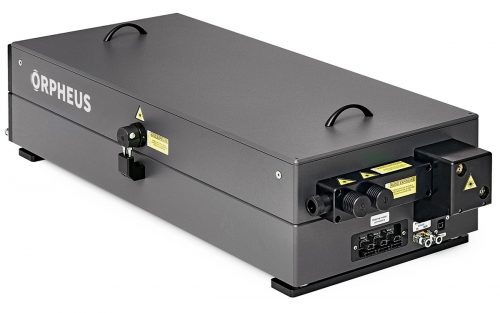
- 结合共线和非共线 OPA 的最佳特性
- 650 – 900 nm & 1200 – 2500 nm 可调波长
- 单脉冲 – 2 MHz 重复频率
- 脉宽 < 100 fs
- 可调光谱带宽
- 波长调谐无间隙的长脉冲模式
- 两个既同步又独立的输出
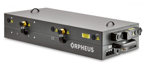
- 210 nm – 16 000 nm 可调波长
- 单脉冲 – 2 MHz 重复频率
- 高达 60 W,0.5 mJ 的泵浦
- 结构紧凑,性价比高
- 选配 CEP 稳定功能
- 11 Mhz、40 Mhz 或 76 Mhz 的重复频率
- <50 fs 的脉宽
- 最高可达 20 W 的高功率型号
- 最高可达 0.6 µJ 的高能量型号
- 高输出稳定性的工业级设计
- CEP 稳定或重复频率锁定

- 100 fs – 20 ps 连续可调脉宽
- 最大单脉冲能量 4 mJ
- 最小脉宽输出 < 100 fs
- POD 和 BiBurst 功能
- 高达 5 次谐波或可调谐扩展
- CEP 稳定或重复频率锁定
- 热稳定性和密封设计
- 190 fs – 20 ps 连续可调脉宽
- 最大输出 1 mJ @ 120 W 或 2 mJ @ 80 W
- 单脉冲 – 2 MHz 重复频率
- POD 和 BiBurst 功能
- 高达 5 次谐波或可调谐扩展
- 风冷型号
- 紧凑的工业级设计
Effect of tissue fixation on the optical properties of structural components assessed by non-linear microscopy imaging
M. A. Markus, D. P. Ferrari, F. Alves, and F. Ramos‑Gomes, Biomed. Opt. Express 8 (14), 3988-4002 (2023).
We need to talk about laser pulse energy stability
L. Kontenis, M. Urbšas, J. Berzinš, and K. Neimontas, in Multiphoton Microscopy in the Biomedical Sciences XXIII, A. Periasamy, P. T. C. So et al., eds. (SPIE, 2023), pp. PC1238410.
Deep tissue multi-photon imaging using adaptive optics with direct focus sensing and shaping
Z. Qin, Z. She, C. Chen, W. Wu, J. K. Y. Lau, N. Y. Ip, and J. Y. Qu, Nature Biotechnology (2022).
Ketogenic diet uncovers differential metabolic plasticity of brain cells
T. Düking, L. Spieth, S. A. Berghoff, L. Piepkorn, A. M. Schmidke, M. Mitkovski, N. Kannaiyan, L. Hosang, P. Scholz, A. H. Shaib et al., Science Advances 37 (8) (2022).
Machine learning-enabled cancer diagnostics with widefield polarimetric second-harmonic generation microscopy
K. Mirsanaye, L. U. Castaño, Y. Kamaliddin, A. Golaraei, R. Augulis, L. Kontenis, S. J. Done, E. Žurauskas, V. Stambolic, B. C. Wilson et al., Scientific Reports 1 (12) (2022).
Biodegradable Harmonophores for Targeted High-Resolution In Vivo Tumor Imaging
A. Y. Sonay, K. Kalyviotis, S. Yaganoglu, A. Unsal, M. Konantz, C. Teulon, I. Lieberwirth, S. Sieber, S. Jiang, S. Behzadi et al., ACS Nano 3 (15), 4144-4154 (2021).
Direct focus sensing and shaping for high-resolution multi-photon imaging in deep tissue
Z. Qin, Z. She, C. Chen, W. Wu, J. K. Y. Lau, N. Y. Ip, and J. Y. Qu, (2021).
Exploring two-photon optogenetics beyond 1100~nm for specific and effective all-optical physiology
T. Fu, I. Arnoux, J. Döring, H. Backhaus, H. Watari, I. Stasevicius, W. Fan, and A. Stroh, iScience 3 (24), 102184 (2021).
Fast optical recording of neuronal activity by three-dimensional custom-access serial holography
W. Akemann, S. Wolf, V. Villette, B. Mathieu, A. Tangara, J. Fodor, C. Ventalon, J. Léger, S. Dieudonné, and L. Bourdieu, Nature Methods 1 (19), 100-110 (2021).
In-vivo tracking of harmonic nanoparticles: a study based on a TIGER widefield microscope [Invited]
L. Vittadello, C. Kijatkin, J. Klenen, D. Dzikonski, K. Kömpe, C. Meyer, A. Paululat, and M. Imlau, Optical Materials Express 7 (11), 1953 (2021).