
Transmission electron microscopy (TEM) is one of the most powerful imaging techniques. Currently, the TEM enables imaging of three-dimensional (3D) structures at the atomic scale. However, the temporal resolution of TEM is often limited by the recording rate of the imaging device used. Thus, to overcome such a limit, a pulsed laser source is applied to trigger the photoemission and, subsequently, to obtain a higher temporal resolution.
The ultrafast electron microscopy (UEM or UTEM) is, essentially, a pump-probe technique where an ultrafast laser pump pulse excites the material and a delayed probe – electron pulse – detects the response at a specific time, correlated with the pump pulse. Accordingly, then the temporal resolution is no longer limited by the speed of the electron detector as it is determined by both the pump laser pulse duration and probe electron pulse. Therefore, the generation of short electron pulses is also quite important and is often carried out using a second or third harmonic of the fundamental frequency of the same ultrafast laser. Furthermore, the UEM requires the laser output with a high pulse repetition rate and high output stability to preserve a high signal-to-noise ratio (SNR).
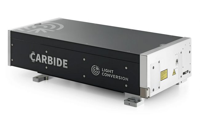
The PHAROS and CARBIDE laser series with femtosecond pulse duration, high repetition rate, and high output stability are excellent sources for ultrafast electron microscopy, making it another promising ultrafast technology.

- 100 fs – 20 ps 连续可调脉宽
- 最大单脉冲能量 4 mJ
- 最小脉宽输出 < 100 fs
- POD 和 BiBurst 功能
- 高达 5 次谐波或可调谐扩展
- CEP 稳定或重复频率锁定
- 热稳定性和密封设计
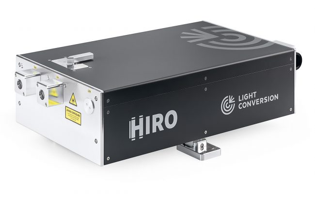
- 190 fs – 20 ps 连续可调脉宽
- 最大输出 1 mJ @ 120 W 或 2 mJ @ 80 W
- 单脉冲 – 2 MHz 重复频率
- POD 和 BiBurst 功能
- 高达 5 次谐波或可调谐扩展
- 风冷型号
- 紧凑的工业级设计
Imaging Nanometer Phonon Softening at Crystal Surface Steps with 4D Ultrafast Electron Microscopy
Y. Zhang, and D. J. Flannigan, (2021).
UEMtomaton: A Source-Available Platform to Aid in Start-up of Ultrafast Electron Microscopy Labs
D. X. Du, S. A. Reisbick, and D. J. Flannigan, Ultramicroscopy 223, 113235 (2021).
Unraveling the Ultrafast Hot Electron Dynamics in Semiconductor Nanowires
L. Wittenbecher, E. V. Boström, J. Vogelsang, S. Lehman, K. A. Dick, C. Verdozzi, D. Zigmantas, and A. Mikkelsen, ACS Nano 1 (15), 1133-1144 (2021).
Characterization of a time-resolved electron microscope with a Schottky field emission gun
P. K. Olshin, M. Drabbels, and U. J. Lorenz, Structural Dynamics 5 (7), 054304 (2020).
Coherent interaction between free electrons and a photonic cavity
K. Wang, R. Dahan, M. Shentcis, Y. Kauffmann, A. B. Hayun, O. Reinhardt, S. Tsesses, and I. Kaminer, Nature 7810 (582), 50-54 (2020).
Development of analytical ultrafast transmission electron microscopy based on laser-driven Schottky field emission
C. Zhu, D. Zheng, H. Wang, M. Zhang, Z. Li, S. Sun, P. Xu, H. Tian, Z. Li, H. Yang et al., Ultramicroscopy 209, 112887 (2020).
High-resolution analogue of time-domain phonon spectroscopy in the transmission electron microscope
E. J. VandenBussche, and D. J. Flannigan, Philosophical Transactions of the Royal Society A: Mathematical, Physical and Engineering Sciences 2186 (378), 20190598 (2020).
Influence of Discrete Defects on Observed Acoustic–Phonon Dynamics in Layered Materials Probed with Ultrafast Electron Microscopy
S. A. Reisbick, Y. Zhang, and D. J. Flannigan, The Journal of Physical Chemistry A 9 (124), 1877-1884 (2020).
Mitigating Damage to Hybrid Perovskites Using Pulsed-Beam TEM
E. J. VandenBussche, C. P. Clark, R. J. Holmes, and D. J. Flannigan, ACS Omega 49 (5), 31867-31871 (2020).
Nanoscale Imaging of Unusual Photoacoustic Waves in Thin Flake VTe2
A. Nakamura, T. Shimojima, Y. Chiashi, M. Kamitani, H. Sakai, S. Ishiwata, H. Li, and K. Ishizaka, Nano Letters 7 (20), 4932-4938 (2020).